Chemistry Behind PET Scans
- Home
- Chemistry Understood
- Chemistry Behind PET Scans


For every PET scan that needs to be performed, there is always a “project” that needs to be conducted before which entails of constructing radionucleotides that are tailored to each specific patient who will consume the radioactive tracer.
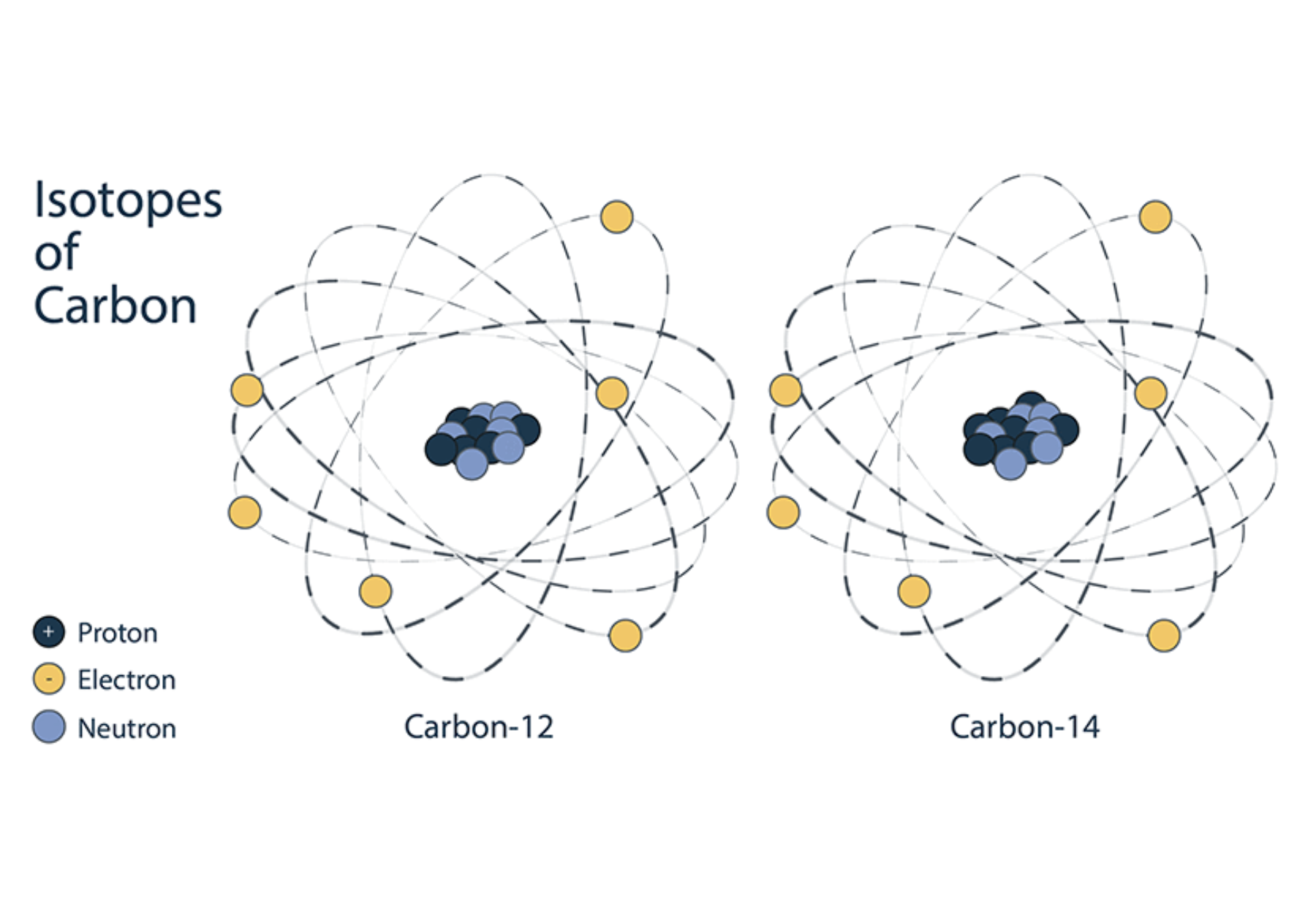
Initially, the process begins with a machine called a cyclotron, which is used to smash high-speed particles together to form radioactive atoms (radionucleotides). The tracers are biological molecules such as proteins, sugars or hormones which are tagged with the radioactive atoms such as Carbon-11, Fluorine-18, Oxygen-15, or Nitrogen-13 (Isotopes of common elements) created in the cyclotron.
When an element has an imbalance in protons and electrons, the isotope will want to try and transform itself into a more stable form, which is resultant in the release of radioactivity (radioactive-decay). Therefore these atoms are radioactive isotopes with short half lives (Interval of time required for one-half of the atomic nuclei of a radioactive sample to decay) and as these radioactive atoms start giving off their radioactivity almost immediately, the tracers need to be made quickly.
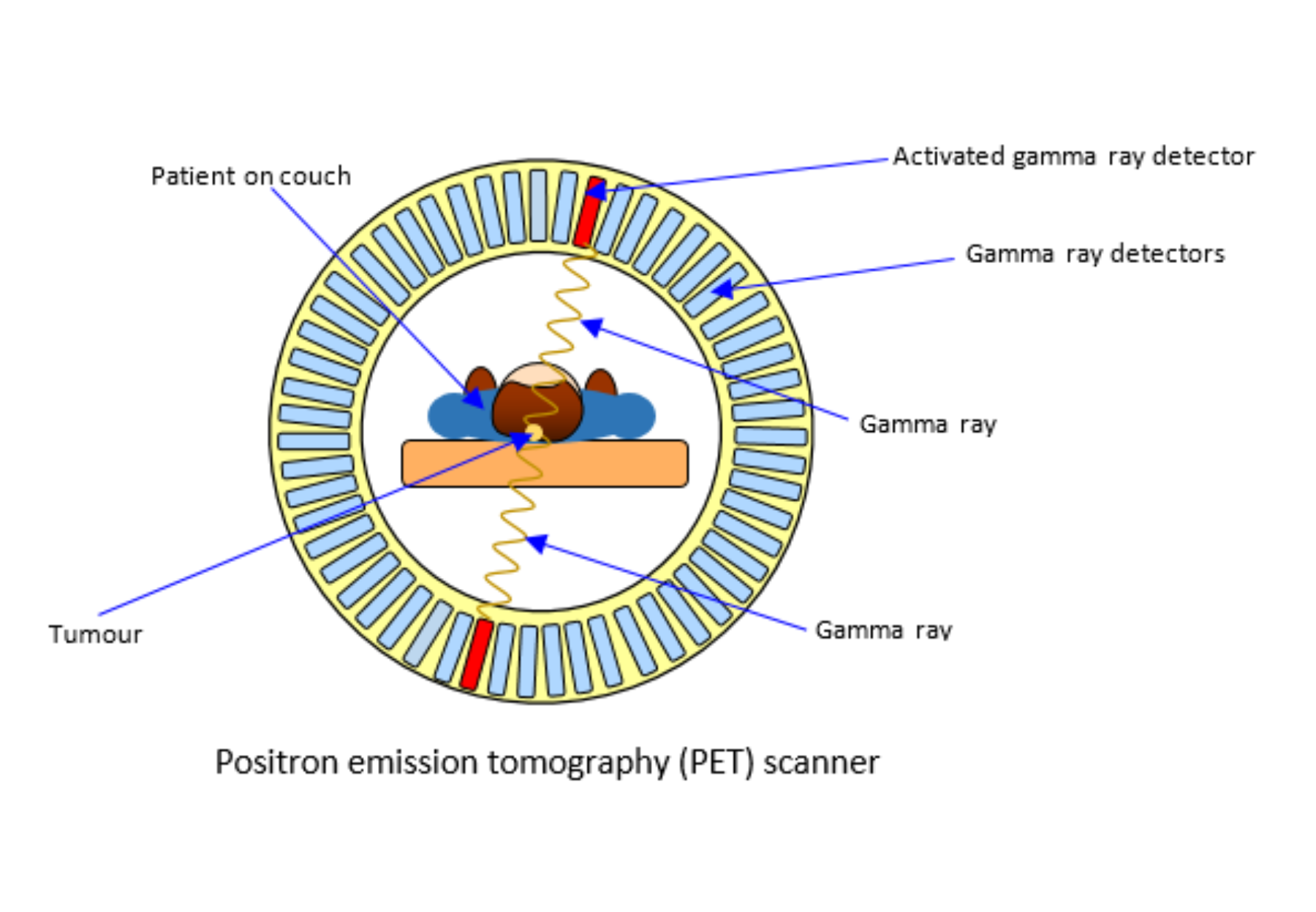
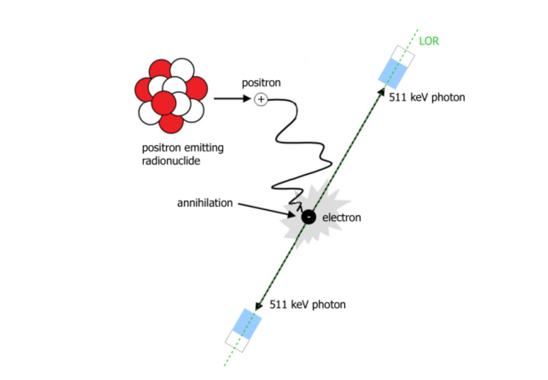
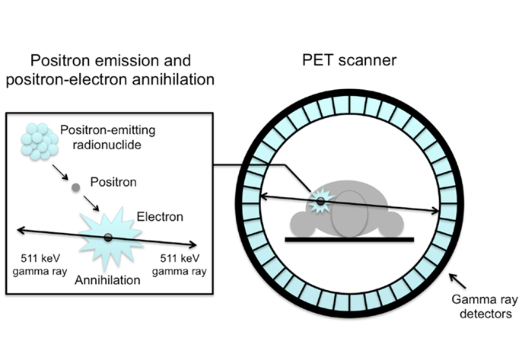
The tracers are usually then injected through an IV (intravenously), which is when the reaction will begin to take place. The radioactive atoms on the tracers start to lose radioactivity giving off subatomic particles called positrons (positively-charged electrons). These positrons are electrons similar to the negatively charged electrons found on all atoms. For the reaction to take place, the positron must hit a negatively charged electron at a precise speed for them to combine and destroy one another (called annihilation); once this occurs, 2 photons are created, these gamma rays travel outside the body in opposite directions. The PET scanner is like a big ring of geiger counters, once two gamma rays on opposite sides are detected, doctors track the location of the tracer and by detecting thousands of rays per second, the patients brain activity is revealed in three-dimension.